info@footandankleunit.co.za | +27 11 647 3735 | +27 11 485 1974 | +27 11 647 3435
PATHOLOGIES
ANKLE CONDITIONS
Osteochondral Lesions
What is it?
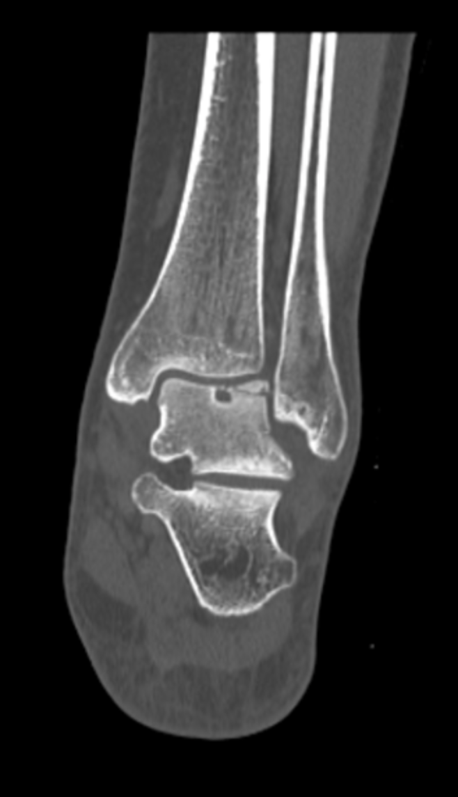
Osteochondral lesions (OCD) refer to damage to the articular cartilage (the smooth tissue that covers the bones in joints) and the underlying bone in the foot and ankle. This condition often affects the talus (the bone that connects the foot to the ankle) and can cause pain, swelling, and joint instability.
The damage typically results from trauma, repetitive stress, or poor blood supply to the bone, leading to a defect where the cartilage and bone begin to deteriorate. In some cases, the cartilage and bone fragment, leading to the creation of a loose piece inside the joint.
CAUSES
Osteochondral lesions can occur due to a variety of reasons, including:
- Trauma or injury: A sudden injury, such as an ankle sprain or fracture, can cause damage to the cartilage and bone, leading to an osteochondral defect. High-impact activities like running or jumping can also contribute to the development of OCD.
- Repetitive stress: Overuse or repetitive activities that put stress on the joint can wear away cartilage and bone over time, increasing the risk of OCD, especially in athletes or individuals who engage in high-impact sports.
- Poor blood supply: In some cases, the bone beneath the cartilage may have a compromised blood supply, leading to poor healing and subsequent development of an osteochondral defect.
- Genetic factors: There may also be a genetic predisposition in some individuals, making them more susceptible to cartilage and bone damage.
- Underlying joint conditions: Conditions like osteoarthritis or rheumatoid arthritis can increase the likelihood of developing osteochondral lesions by causing degenerative changes to the joint over time.
SYMPTOMS
Symptoms of osteochondral lesions can vary depending on the severity and location of the damage, but common signs include:
- Pain: Pain in the affected joint, especially during weight-bearing activities such as walking or running, is common. The pain may be localized to the area of the defect (usually the ankle or foot), although often the locale of the pain is vague
- Swelling: The joint may appear swollen, particularly after physical activity or injury.
- Stiffness: The affected foot or ankle may feel stiff, especially after long periods of inactivity or in the morning.
- Locking or catching sensation: If the defect results in loose bone or cartilage fragments, the joint may experience a locking or catching sensation during movement, making it difficult to move the joint smoothly.
- Instability: In more severe cases, the joint may feel unstable or weak due to the damage to the cartilage and underlying bone.
DIAGNOSIS
To diagnose osteochondral lesions, a healthcare provider will perform a combination of physical exams and imaging studies:
- Physical examination: The doctor will assess the joint for tenderness, swelling, and restricted range of motion. Special tests may be performed to evaluate joint function and pinpoint the source of pain.
- X-rays: X-rays can help identify bone defects, fractures, or signs of joint degeneration associated with osteochondral lesions. However, cartilage damage may not always be visible on X-rays.
- MRI: Magnetic Resonance Imaging (MRI) is the most accurate imaging technique for detecting osteochondral lesions. It provides detailed images of both the cartilage and bone, helping to determine the size, location, and severity of the defect.
- CT scan: In some cases, a CT scan may be used for detailed visualization of the bone and cartilage, especially if the defect is complex or located in a hard-to-see area.
- Arthroscopy: Arthroscopy (a minimally invasive procedure where a small camera is inserted into the joint) may be used to directly visualize the defect, confirm its size, and assess the joint for further damage.
TREATMENT OPTIONS
Non-Surgical Treatment:
For mild to moderate osteochondral lesions or for patients who are not experiencing significant symptoms, non-surgical treatment may be effective:
- Rest and activity modification: Resting the affected joint and avoiding high-impact activities or sports can help reduce stress on the joint and promote healing. Low-impact exercises like swimming or cycling may be recommended.
- Physical therapy: Can help improve range of motion, strengthen the muscles around the joint, and reduce pain and stiffness. Exercises may focus on improving the overall function of the foot and ankle.
- Nonsteroidal anti-inflammatory drugs (NSAIDs): NSAIDs, such as ibuprofen, may be used to reduce inflammation, pain, and swelling in the joint.
- Bracing or orthotics: A custom brace or orthotics can help offload pressure from the affected joint, provide support, and prevent further joint damage.
Surgical Treatment:
In more severe cases or when conservative treatments fail, surgery may be required to repair or remove the damaged cartilage and bone. Surgical options include:
- Arthroscopic debridement: A minimally invasive procedure to remove any damaged cartilage and bone fragments from the joint. This can help alleviate symptoms and prevent further degeneration of the joint.
- Microfracture surgery: This procedure involves creating tiny holes in the bone beneath the cartilage to stimulate bone healing and the growth of new cartilage. It is typically used for small to moderate defects.
- Osteochondral autograft transplantation: In this procedure, healthy cartilage and bone are taken from a non-weight-bearing area of the joint and transplanted to the defect site.
- Osteochondral allograft transplantation: For larger defects, cadaveric cartilage and bone may be transplanted to repair the damage.
- Platelet Rich Plasma/Stem cell therapy: In some cases, platelet rich plasma/stem cell injections may be used to promote cartilage regeneration and improve healing in the defect area.
- Joint fusion: In very severe cases, where the joint is extensively damaged, joint fusion may be performed to stabilize the joint and relieve pain.
PROGNOSIS
The prognosis for osteochondral lesions depends on the size, location, and severity of the defect, as well as the treatment approach:
- Mild defects: With appropriate non-surgical treatment and rest, many people with small defects can experience significant improvement and return to normal activity.
- Severe defects: Larger defects, especially those that cause joint instability or locking, may require surgery to restore function. Surgical outcomes are generally good, with many patients experiencing pain relief and improved mobility.
- Chronic issues: If left untreated, osteochondral lesions can lead to long-term joint pain, degeneration, and even osteoarthritis in the affected joint.