info@footandankleunit.co.za | +27 11 647 3735 | +27 11 485 1974 | +27 11 647 3435
PATHOLOGIES
FRACTURES AND TRAUMA
Calcaneus Fracture
What is it?
A calcaneus fracture refers to a break in the calcaneus bone, which is the large bone that forms the heel of the foot. This type of fracture is commonly caused by high-impact trauma, such as a fall from a height or a car accident. The calcaneus is one of the most important weight-bearing bones in the foot, and fractures can significantly affect mobility and function.
Calcaneus fractures can vary in severity, from simple cracks to complex fractures where the bone is shattered into multiple pieces. These fractures are often associated with other injuries to the foot or lower extremity.
Types
Calcaneus fractures are classified based on the location of the break and whether the fracture is displaced:
- Non-displaced fracture: The bone cracks but remains in alignment.
- Displaced fracture: The broken pieces of bone are out of alignment.
- Comminuted fracture: The calcaneus is broken into several pieces.
- Intra-articular fracture: The fracture extends into the subtalar joint (the joint below the ankle), which can lead to joint instability and long-term arthritis.
- Stress fracture: A small crack in the bone caused by repetitive stress or overuse.
CAUSES
The most common causes of calcaneus fractures include:
- Falls from a height: Falling from a height (such as off a ladder, scaffolding, or during a jump) is a primary cause of calcaneus fractures, as the bone is directly impacted.
- Car accidents: A direct blow to the heel during a motor vehicle accident can cause a calcaneus fracture.
- Sports injuries: High-impact sports (e.g., soccer, skiing, or football) can also lead to calcaneus fractures, often from sudden twisting motions or jumps.
- Repetitive stress: In rare cases, repetitive stress on the calcaneus (often seen in athletes or military personnel) can cause stress fractures over time.
SYMPTOMS
Common symptoms of a calcaneus fracture include:
- Pain: Intense pain in the heel or back of the foot, especially when trying to stand or walk.
- Swelling and bruising: Significant swelling around the heel, and bruising may appear on the foot or ankle.
- Deformity: A noticeable deformity in the heel, especially in cases of severe fractures.
- Inability to walk: Difficulty or inability to bear weight on the affected foot due to pain and instability.
- Tenderness: Extreme tenderness around the heel bone when touched.
DIAGNOSIS
- Physical examination: A healthcare provider will assess the alignment of the foot and check for areas of tenderness, deformity, and limited movement.
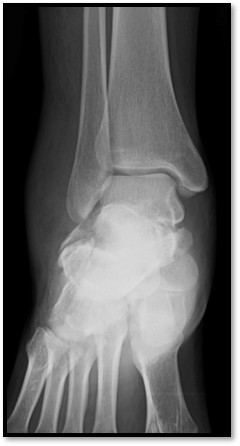
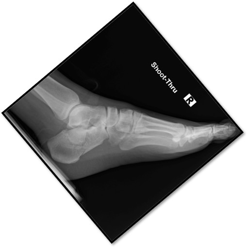
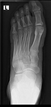
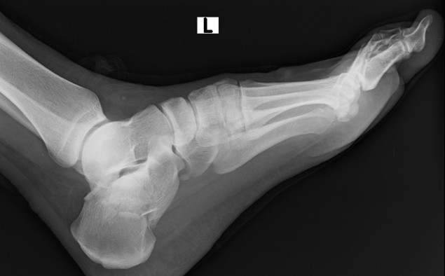
- X-rays: X-ray imaging is the primary diagnostic tool for calcaneus fractures. Multiple views (e.g., lateral and axial) are typically taken to determine the type of fracture and its location.
- CT scan: In complex fractures, a CT scan can be used to get a more detailed view of the bone fragments and joint involvement.
- MRI: If there is concern about soft tissue damage, such as ligaments or cartilage injury, an MRI may be ordered.
TREATMENT OPTIONS
Non-Surgical Treatment:
For non-displaced fractures or fractures with minimal displacement, the treatment approach typically includes:
- Rest and immobilization: The foot is usually placed in a cast or boot to immobilize the area while it heals. Weight-bearing is avoided for several weeks.
- Ice and elevation: Applying ice to reduce swelling and elevating the foot to minimize inflammation is helpful in the early stages.
- Pain management: Over-the-counter pain relievers, such as NSAIDs, can help control pain and reduce swelling.
- Physical therapy: Once healing has occurred, physical therapy is important to regain strength, flexibility, and function in the foot and ankle.
Surgical Treatment:
For displaced fractures, fractures that involve the subtalar joint, or comminuted fractures, surgery may be required to properly realign the bones and prevent complications:
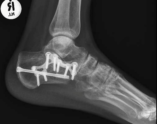
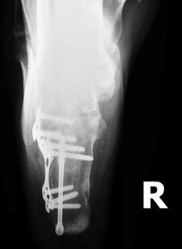
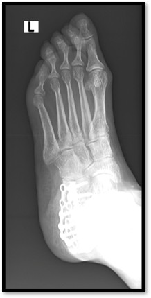
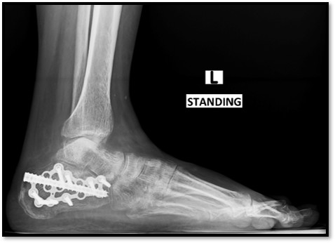
- Open reduction and internal fixation (ORIF): In this procedure, the broken bones are realigned and secured using plates, screws, or pins. This is the most common surgical method for calcaneus fractures.
- Fusion: In cases where there is significant joint damage or arthritis, a subtalar joint fusion may be performed to stabilize the joint.
- Excision of fragments: In some cases, small bone fragments that are causing pain or hindering movement may be surgically removed.
RECOVERY AND REHABILITATION
- Initial recovery: After surgery or non-surgical treatment, the foot is generally immobilized for 6-8 weeks to allow the bones to heal. During this time, weight-bearing is restricted.
- Physical therapy: Rehabilitation is crucial after a calcaneus fracture, especially if surgery was involved. Therapy focuses on restoring mobility, strength, and function to the foot and ankle.
- Gradual return to activity: Full recovery can take several months. High-impact activities such as running or jumping may need to be avoided for 6-12 months, depending on the severity of the injury.
- Complications: In some cases, calcaneus fractures can lead to long-term complications such as arthritis in the subtalar joint, chronic pain, or limited motion in the foot.
PROGNOSIS
- Good recovery with appropriate treatment: Most people recover well from a calcaneus fracture, particularly with early diagnosis and treatment. Full recovery can take several months, with gradual return to normal activity.
- Possible complications: If not treated promptly or correctly, calcaneus fractures can result in chronic pain, deformity, or arthritis in the subtalar joint, particularly in intra-articular fractures.
- Surgical intervention: Surgery can significantly improve outcomes, especially for displaced fractures, by restoring bone alignment and joint stability.