PATHOLOGIES

MIDFOOT AND HINDFOOT CONDITIONS

Base of 5th Metatarsal Fracture

What is it?

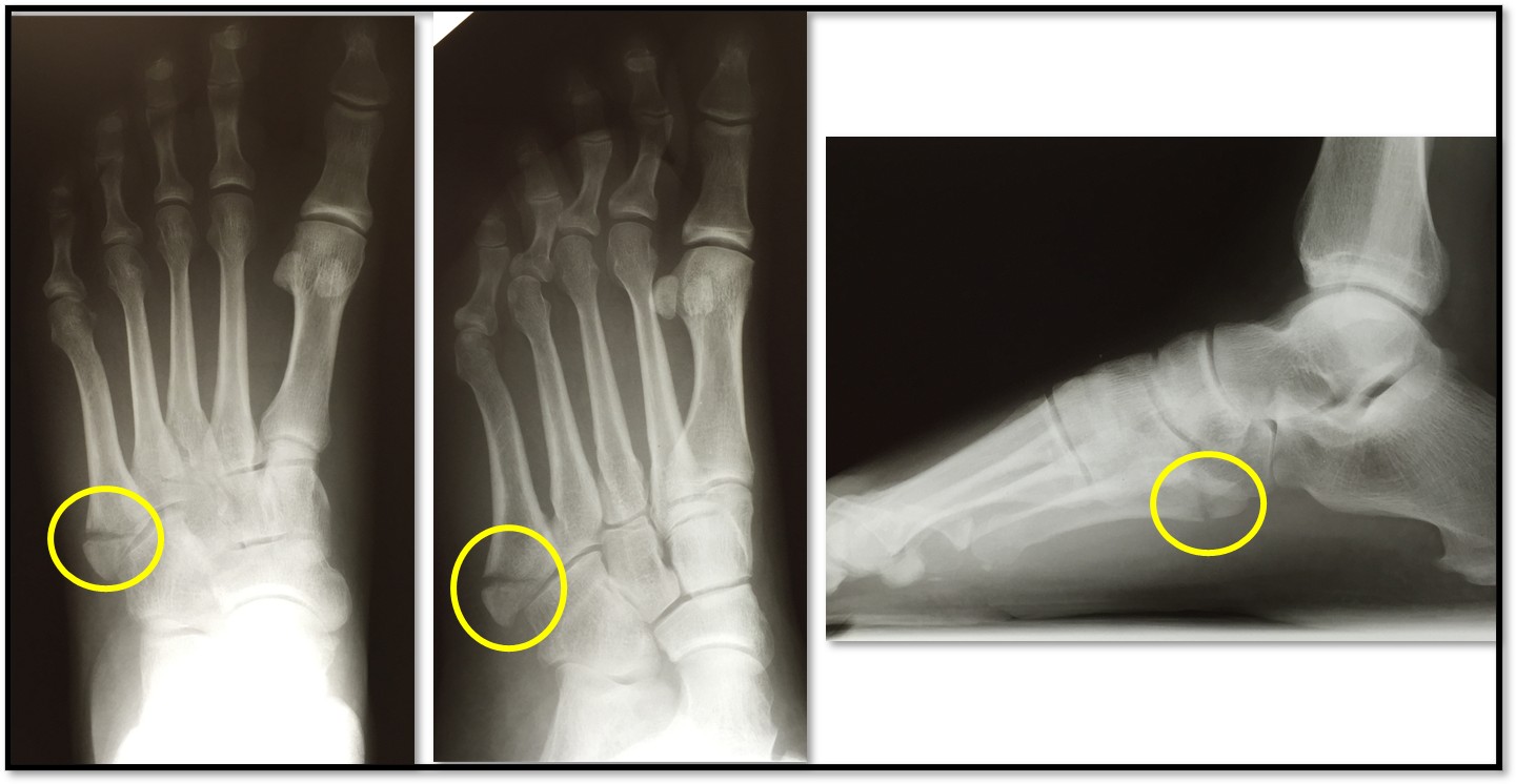
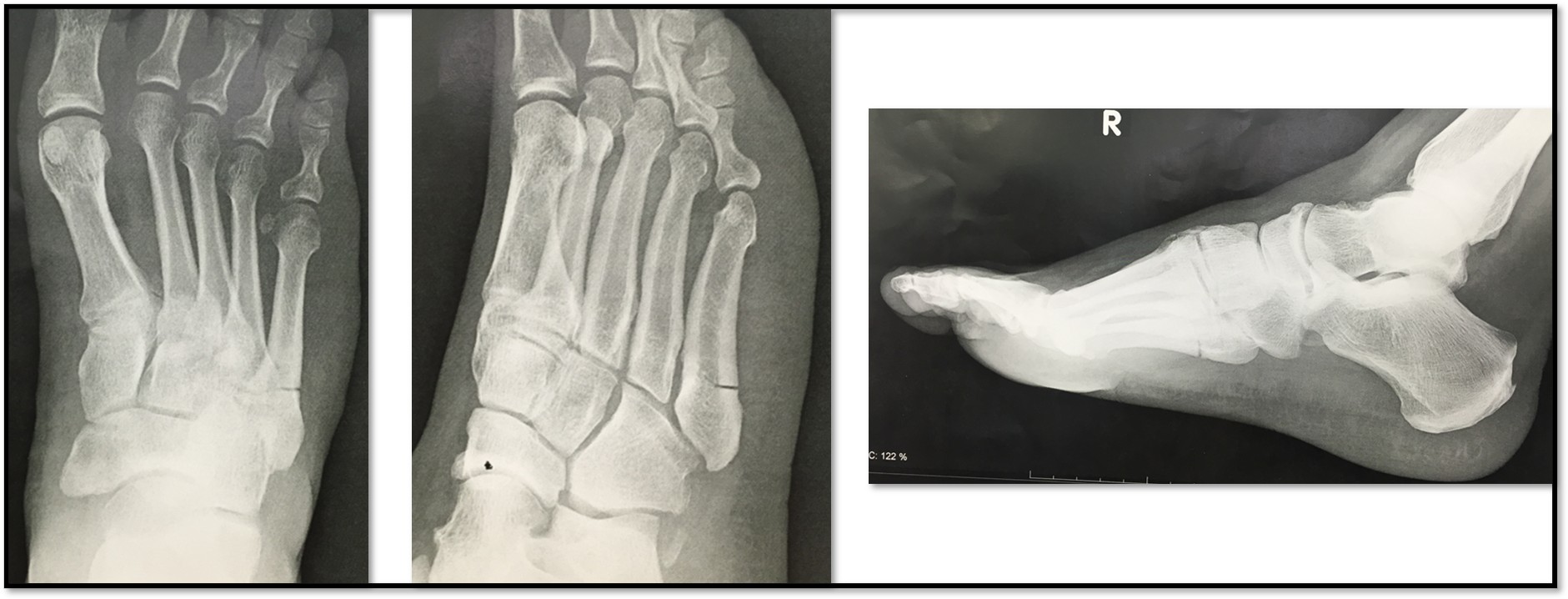
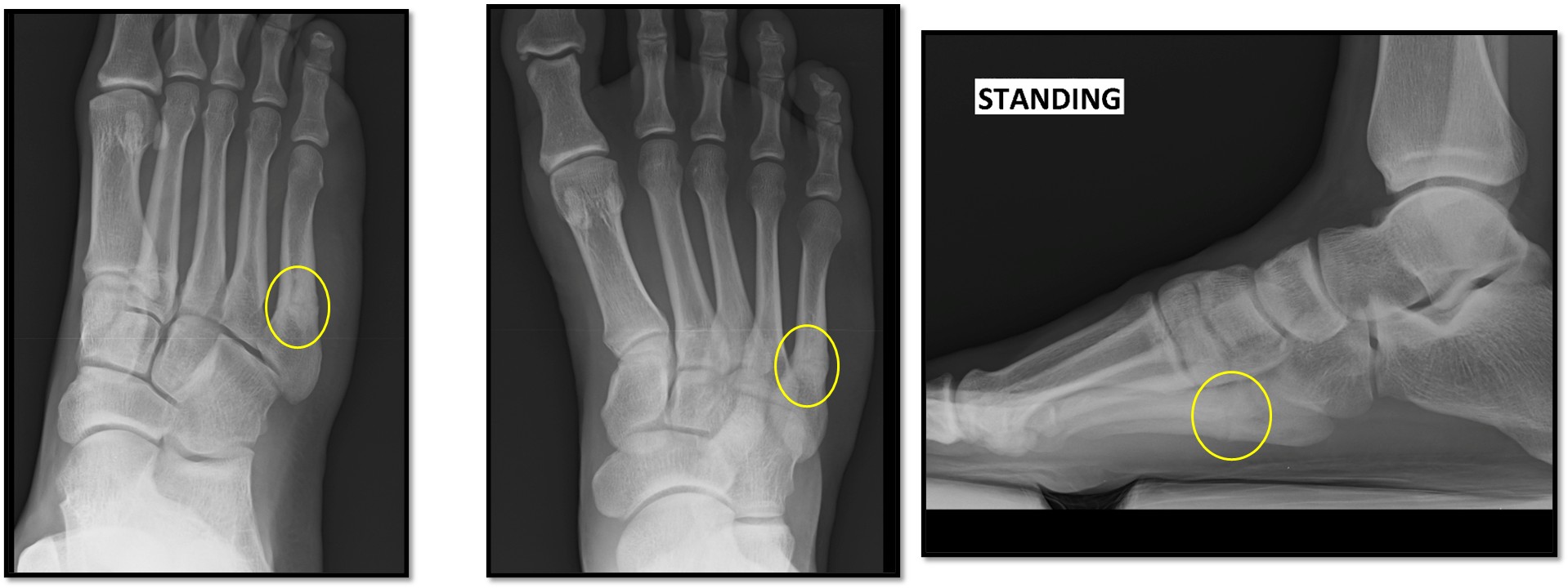
A base of 5th metatarsal fracture is a break at the outer side of the foot, where the fifth metatarsal (the long bone leading to the little toe) meets the midfoot. These fractures are common and vary by location and mechanism:

  1. Avulsion fracture
    • A small bone fragment is pulled off by the peroneus brevis tendon or lateral band of the plantar fascia.
    • Often occurs with an ankle inversion injury (“rolled ankle”).
  2. Jones fracture
    • Occurs in the metaphyseal–diaphyseal junction, about 1.5–3 cm from the base.
    • Mechanism: acute adduction force on a plantarflexed foot or repetitive stress.
    • Higher risk of delayed healing or nonunion due to relatively poor blood supply.
  3. Stress fracture
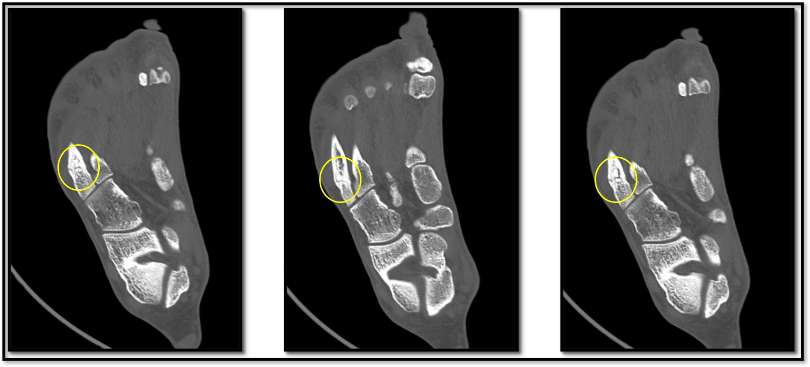
    • A hairline crack in the proximal diaphysis from overuse (e.g., running, dancing).
    • Presents with gradual onset of lateral foot pain.

Types

Calcaneus fractures are classified based on the location of the break and whether the fracture is displaced:

  1. Non-displaced fracture: The bone cracks but remains in alignment.
  2. Displaced fracture: The broken pieces of bone are out of alignment.
  3. Comminuted fracture: The calcaneus is broken into several pieces.
  4. Intra-articular fracture: The fracture extends into the subtalar joint (the joint below the ankle), which can lead to joint instability and long-term arthritis.
  5. Stress fracture: A small crack in the bone caused by repetitive stress or overuse.
CAUSES AND RISK FACTORS
  • Trauma: Inversion sprains or direct impact.
  • Repetitive stress: Especially in athletes (runners, basketball players).
  • Foot structure: Cavus (high-arched) feet may transfer more load laterally.
  • Poor conditioning: Sudden increases in activity without gradual buildup.
  • Localized pain at the outer mid-foot worsens with weight-bearing.
  • Tenderness to touch at the base of the 5th metatarsal.
  • Swelling and bruising over the fracture site.
  • Difficulty walking or pushing off the little toe.
  • Clinical exam: Point tenderness, swelling, and possible pain on resisted eversion.
  • X-rays: Essential to classify fracture type (avulsion vs. Jones vs. stress).
  • MRI/CT: May be used if healing problems arise or to assess stress injuries.

Non-Surgical

  • Avulsion fractures often heal well with:
    • Protected weight-bearing in a stiff-soled shoe or boot for 4–6 weeks.
    • Ice, NSAIDs, and early motion within comfort.
  • Jones and stress fractures:
    • Non-weight-bearing cast or boot for 6–8 weeks, followed by gradual return.
    • Low-impact cross-training (swimming, cycling) during immobilization.

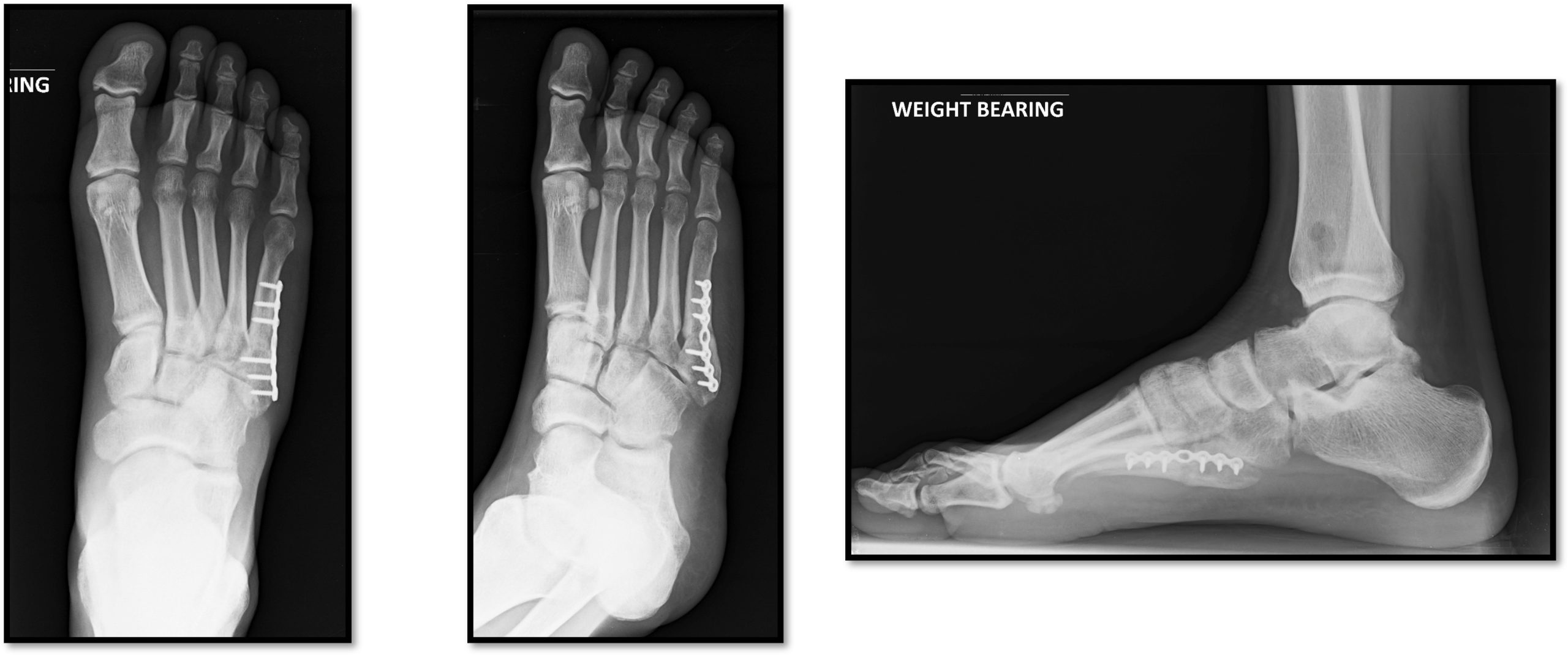
Surgical

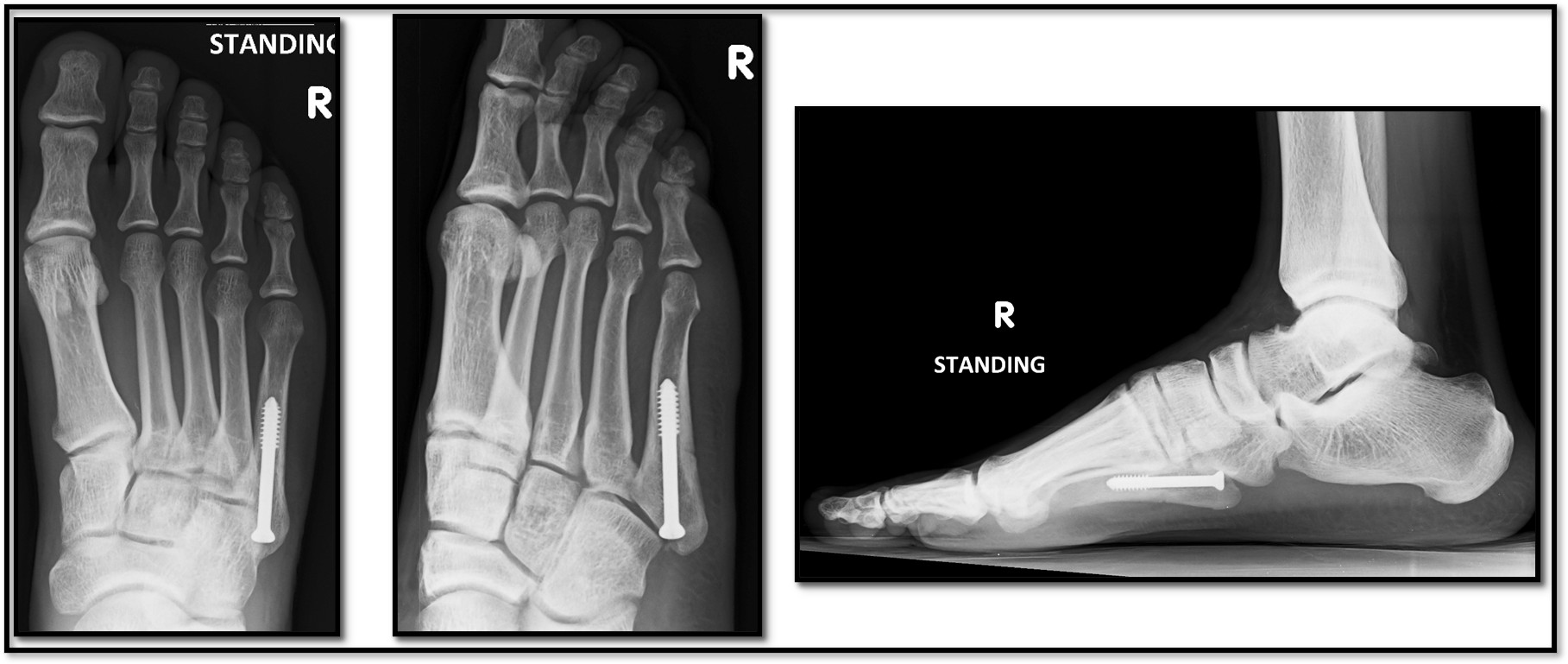
  • Considered for Jones fractures in athletes or high-risk patients to reduce nonunion risk.
  • Plate and screws or Intramedullary screw fixation are most common methods
  • Post-op: boot immobilization and non-weight-bearing for 6 weeks, then progressive rehab.
  1. Avulsion fractures: Excellent healing in most cases within 6–8 weeks.
  2. Jones/stress fractures: Longer healing times; 80–90% union rate with non-surgical care, >95% with surgery in athletes.
  3. Nonunion risk is higher in Jones fractures—monitor healing via imaging.